Image Details
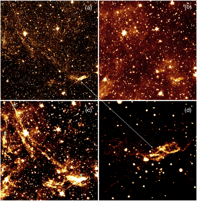
Caption: Figure 7.
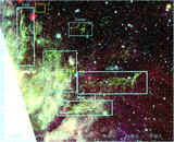
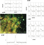
Shock-excited molecular hydrogen in the southeast region of the HB 3 SNR (see Figure 5 and the exact location is marked as a white box). The diffuse emission in the WIRC H2 2.12 μm image (a) ranges 0.179–2.4 MJy sr−1 (∼1.91–26.7 × 10⁻⁶ erg s−1 cm−2 sr−1). The emission in Spitzer 3.6 and 4.5 μm imaging, upper right and lower left (b, c), respectively, ranges 0.41–0.91 (typical background is 0.35) MJy sr−1 and 0.25–1.28 (0.2) MJy sr−1, respectively. The images (a)–(c) are centered on R.A. 2h19m59.6ˢ and decl. +62°06′39.1″ (J2000) with an FOV of 8.8′ x 9.2′. The narrow-filter WIRC image (d) zoomed on the brightest emission shows complex shock structures, and is centered on R.A. 2h19m59.6ˢ and decl. +62° 06′ 39.1″ (J2000) with an FOV of 78″ × 73″. The filament positions of A, B, and C on the image (c) (see Table 4) are marked.
Other Images in This Article

Show More
Copyright and Terms & Conditions
© 2021. The American Astronomical Society. All rights reserved.