Image Details
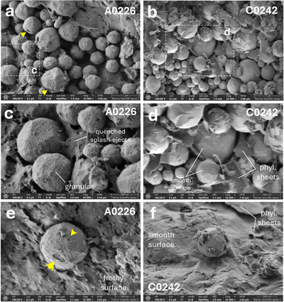
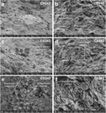
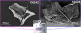
Caption: Figure 3.
Electron microscope images in secondary electron (SE) mode of magnetite grains in A0226 and C0242 particle surfaces. (a) Panoramic SE image of framboidal magnetite on the A0226 surface, showing micro- to nano-impact craters (highlighted by yellow arrows), granular surface morphology, and magnetite grains interconnected by a “cobweb” texture. (b) Panoramic SE image of framboidal magnetite on the C0242 surface, showing a pristine, smooth morphology commonly associated with phyllosilicate (phyl.) sheets. (c) High-magnification view of the area in panel (a), highlighting quenched splash ejecta feature on weathered magnetite. (d) High-magnification view of the area in panel (b), showing a smooth magnetite surface and associated phyllosilicate sheets. (e) Individual magnetite grain in A0226 exhibiting nanometer impact craters on a frothy surface. (f) Individual magnetite grain in C0242 showing a smooth surface on a phyllosilicate-rich matrix.
Other Images in This Article
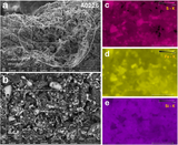
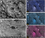
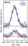
Show More
Copyright and Terms & Conditions
© 2026. The Author(s). Published by the American Astronomical Society.